Skin, nail and hair for mycology (dermatophyte fungal fungus fungi tinea ringworm onychomycosis superficial mycoses pityriasis athlete's foot)
Microbiology
Notes
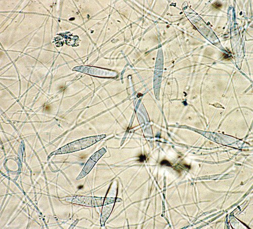
- Routine investigation includes microscopy and culture for dermatophytes, Candida, Malasezzia furfur (pityriasis versicolor) and other moulds
- Pityriasis versicolor is diagnosed solely by microscopy as Malasezzia does not grow on standard laboratory culture media
- A positive microscopy result is diagnostic for fungal infection even in the absence of a positive culture
- In the event of a small sample being received microscopy will be given priority over culture
- The information given here is intended for use by healthcare professionals. Please see Lab Tests Online-UK for more general advice, links and background.
Sample requirements
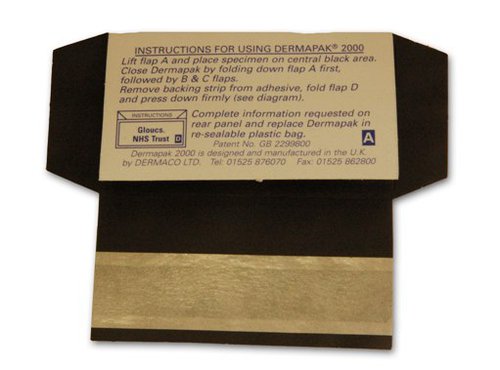
Collect skin scrapings, nail clippings or plucked hair into Dermapak
- The edges of skin lesions yield the greatest quantities of viable fungus. Lesions should be scraped with a blunt scalpel blade.
- Material should be taken from any discoloured, dystrophic or brittle parts of the nail. The affected nail should be cut as far back as possible through the entire thickness and should include any crumbly material.
- When there is superficial involvement (as in white superficial onychomycosis) nail scrapings may be taken with a curette.
- Samples from the scalp should include skin scales and plucked hairs or hair stumps. Cut hairs are not suitable for direct examination.
Required information
- Animal contact
- Site of infection
- Presence of immunosuppression
Storage/transport
Store and transport at room temperature
Turnaround time
Microscopy: up to 5 days
Culture: 2 – 3 weeks